


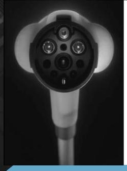
Innovia Multi-nuclear MRI Coils
Built for multi-nuclear MRI.
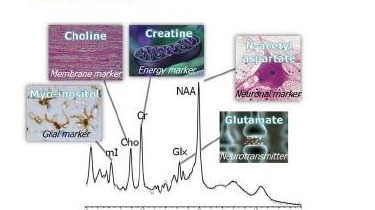
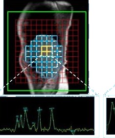
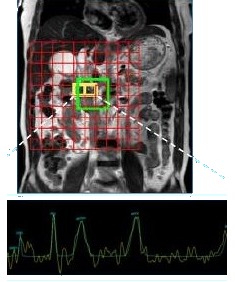
The Multinuclear Resonance Spectrum
A frontier technology for revealing in-vivo molecular metabolism and microenvironment interactions.

Multi-nuclear MRI uses a range of nuclei (such as ¹²⁹Xe, ³¹P, ²³Na, ¹⁹F, ¹³C, ³He, and ²H) to capture in-vivo information about cellular metabolic processes, characterise the interaction between cells and their microenvironment, and reveal how those interactions regulate and remodel the surrounding microstructures.
Multi-nuclear MRI offers high spatial resolution and high specificity, and is currently the only technology capable of in-vivo, non-invasive, quantitative analysis of dynamic molecular metabolism in the human body.
- ●Stable isotopes with no radiation risk
- ●Non-toxic labelling of endogenous metabolites such as glucose, amino acids and fatty acids
- ●Safe, repeatable tracking of metabolic dynamics for both basic research and clinical translation
Nuclide Applications
Non-invasively monitors intracellular high-energy phosphate compounds such as ATP and phosphocreatine, revealing cellular energy state and metabolic dynamics.
Non-invasively measures the distribution and concentration changes of sodium ions inside and outside cells and tissues, reflecting cellular osmotic pressure and microenvironmental function.
In-vivo tracking of deuterium-labelled metabolites for multi-nuclear MRI, enabling quantitative monitoring of dynamic changes in cellular metabolic pathways.
Non-invasively analyses cellular metabolic pathways by labelling carbon-based metabolites, revealing metabolic activity and functional state.
Nuclide Technical Profile
100% naturally abundant and sensitive to phosphorus metabolites, ideal for energy metabolism studies.
The only MRI-active sodium isotope, reflecting cellular and tissue ionic environments.
Spans a wide chemical shift range, enabling non-invasive monitoring of metabolic pathways.
Behaves like ¹H but with broader lines and lower sensitivity, useful for tracing labelled metabolites in vivo.
¹⁹F is rare in the human body and produces almost no background signal, making it a highly sensitive and specific probe for tracking drug distribution and metabolic processes.
Clinical Applications of Multi-Nuclear MRI
Tumour Assessment
Early tumour screening and subtype identification, providing a basis for precision treatment.
Cardiovascular Assessment
Rapidly assesses myocardial function, blood flow, and ion distribution, supporting diagnosis and treatment monitoring of cardiovascular disease.
Neurological Assessment
Detects abnormal brain metabolism and microenvironmental changes, assisting early diagnosis and tracking of neurodegenerative diseases.
Endocrine System Assessment
Non-invasively monitors metabolite levels, providing a reference for diagnosis and treatment evaluation of endocrine disorders such as diabetes.
Digestive System Assessment
Analyses metabolic and ionic dynamics of the liver, intestines, and pancreas, supporting early disease detection and treatment evaluation.
Respiratory System Assessment
Evaluates pulmonary gas exchange, microstructure, and local metabolic changes, supporting diagnosis and monitoring of respiratory diseases.
Innovia’s Current Multi-Nuclear MRI Coil Family
Dual-Tuned X / ¹H Head Coil
- ●Captures both ¹H and X-nucleus images on a single dual-tuned coil, with no coil swap (²³Na, ³¹P, ²H, etc.)
- ●Multi-nuclear scan workflow matches that of routine ¹H imaging
- ●Fully integrated into the clinical workflow
Dual-Tuned X / ¹H Preclinical Coil
- ●Dual-tuned design (Tx / Rx integrated)
- ●Supplied with a dedicated animal bed and scan-positioning markers
- ●Supports a range of nuclei (³¹P, ²H, etc.)
Flex Coil X-140
- ●Integrated transmit-receive (Tx / Rx) design
- ●Supports cross-anatomy imaging research
- ●Improved image SNR and clearer spectroscopy
Coil Extension Matrix
Innovia multi-nuclear MRI coil extension roadmap.
Dual-Tuned X / ¹H Breast Coil
Breast-dedicated coils including ²³Na/¹H, ³¹P/¹H, ²H/¹H, and others.
Dual-Tuned X / ¹H Torso Coil
Body-dedicated coils including ²³Na/¹H, ³¹P/¹H, ²H/¹H, and others.
Advanced Dual-Tuned Coil
Higher channel count, broader coverage, more flexible design.
Dual-Tuned Multi-Nuclear Head Coil
Innovia’s dual-tuned head coil lets you complete a brain examination within a routine scan time, capturing both proton and other-nuclei images (for example ³¹P, ¹³C, ²³Na) without changing coils. A complete brain study can be completed in under 30 minutes; a standalone ²³Na brain scan in 15 minutes.
The workflow is identical to a routine hydrogen-proton scan. The scan card automatically recognises the dual-tuned head coil, and the nucleus type is treated as another scan parameter. Multi-nuclear spectra and image review, and pushing data to PACS, are all fully integrated.
Combined with our multi-nuclear professional package, the head coil seamlessly integrates multi-nuclear research into your daily scanning workflow.

Clinical Examples
³¹P / ²³Na / ²H Channel
- ●Transmit / Receive (Tx/Rx)
- ●Quadrature polarisation
¹H (Proton) Channel
- ●Transmit / Receive (Tx/Rx)
- ●Quadrature polarisation
Features
- ●Dual-frequency integrated transmit-receive design (³¹P/¹H, ²³Na/¹H, ²H/¹H)
- ●Inner diameter: 26.5 cm
- ●Resonator length: 24 cm
- ●Fits 99% of the population
- ●Birdcage circular polarisation excitation, with maximum excitation efficiency and a uniform B1 field
- ●Fixed-tuned open design with an open rear, making it easy to position and secure the patient’s head
- ●Compatible with 3.0T MRI systems from Philips, Siemens, GE and others
- ●Mechanical structure adapted to the scan bed; supplied with mirror and cushioning
- ●Backed by research support for sequence and parameter optimisation
Flex Coil X-140
Self-contained Tx/Rx X-Channel
Unleash full potential.
- ●Plays a key role in early disease detection and precision evaluation
- ●Provides functional and metabolic information for specific tissues
- ●Extends the boundaries of imaging in chronic-disease and functional assessment
Seamless multi-nuclear integration. Unlocking the potential of the whole body.
Non-invasive. In vivo. Targeted.
Preview the demo images
³¹P spectroscopy can detect changes in myocardial energy reserve in heart failure patients before left-ventricular ejection fraction (LVEF) declines, and can identify mitochondrial dysfunction in Alzheimer’s and Parkinson’s disease before structural imaging shows abnormalities. ²³Na imaging can detect abnormal renal sodium reabsorption earlier than estimated glomerular filtration rate (eGFR), and can be correlated with systemic metabolic disorders in diabetic patients.
In oncology, combining ³¹P and ²H allows more accurate and earlier evaluation of glucose and energy metabolism. In sports medicine, ³¹P enables direct, dynamic monitoring of muscle energy supply. In arthritis, ²³Na concentration is closely correlated with early cartilage degeneration and inflammatory activity.
¹⁹F / ¹²⁹Xe imaging can detect localised ventilation deficits in obstructive lung disease earlier than spirometry, with greater data stability. In non-alcoholic fatty liver disease (NAFLD), ³¹P spectroscopy correlates closely with hepatic fibrosis and inflammation severity.
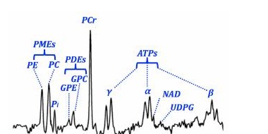
³¹P Spectrum Images
Demonstration spectra captured with the Flex Coil X-140.
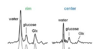
Precisely quantifies hepatic fat and iron content, evaluates hepatic metabolic state and fibrosis, and identifies the metabolic signature of liver tumours.
Evaluates exocrine pancreatic function and inflammation, detects metabolic features of pancreatic tumours, and supports research into diabetes and pancreatic β-cell function.

Detects myocardial energy failure early, evaluates myocardial-cell integrity and diffuse fibrosis, and supports research into myocardial substrate metabolism and ischaemia.
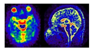
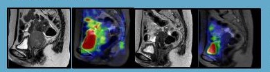
²³Na Images
Demonstration sodium MRI images captured with the Flex Coil X-140.
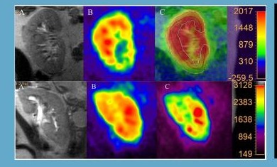
A reference surrogate marker for evaluating renal function and injury, enabling differentiation between pre-renal and renal acute kidney injury.
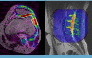
Early diagnosis of cartilage degeneration and osteoarthritis. Proteoglycan loss precedes structural damage and shows up as a reduced local sodium signal.
On ²³Na MRI, the high signal at the tumour site disappears after radiotherapy, indicating cell apoptosis and a fall in intracellular sodium.
Multi-Nuclear Surface Coil
Integrated Tx/Rx
A 14 cm-diameter integrated transmit-receive multi-nuclear flexible surface coil, supporting imaging, spectroscopy and research applications across a range of nuclei, suitable for use anywhere on the body.
Multi-nuclear examinations can be completed within routine scan times, with a workflow identical to standard ¹H imaging. The scan card automatically recognises the coil, and the nucleus is simply one of the scan parameters.
P-140 Coil (³¹P)
Designed for phosphorus (³¹P) spectroscopy and research across all anatomical regions of the whole body. Significantly improves ³¹P signal-to-noise ratio (SNR) and simplifies ³¹P spectral display. Researchers can study muscle metabolic kinetics by tracking changes in the PCr/Pi ratio over time.
Na-140 Coil (²³Na)
Designed for sodium (²³Na) imaging and research across all anatomical regions, completing a sodium (²³Na) knee examination in approximately 15 minutes. Sub-millisecond TE acquisition technology helps capture short-T2 signals.
Dual-Tuned Surface Coil
A dual-tuned surface coil for multi-nuclear MRI applications, designed for versatile research use. Supports imaging and spectroscopy with ³¹P, ²³Na, ¹⁹F, ¹³C, ²H and other nuclei. Operates reliably on 3.0T MRI systems from Philips, Siemens, GE and others, with an effective field of view of S/I 140 mm × R/L 140 mm.
Functional Characteristics
Integrated Transmit-Receive Design
Combines transmit and receive functions to significantly improve local SNR and optimise RF efficiency.
Localised High Sensitivity
A close-contact surface-coil structure that significantly improves SNR in the target region, delivering clearer data for microstructural imaging and metabolic-signal acquisition.
Flexible Multi-Scenario Use
Compatible with all 3.0T systems on the market, suitable for research in oncology, musculoskeletal, neurological and metabolic studies.
Multi-Nuclear Animal (Preclinical) Coil
Dual-Tuned X / ¹H
A multi-nuclear animal coil with a dual-tuned design and integrated transmit-receive, offering excellent RF uniformity and signal stability. Supplied with a dedicated animal bed and scan-positioning marker system, simplifying animal positioning and improving experimental reproducibility.
Supports both hydrogen-nucleus and a range of multi-nuclear applications, including ¹²⁹Xe, ³¹P, ²³Na, ¹⁹F, ¹³C, ³He, and ²H, meeting the needs of multi-nuclear imaging and spectroscopy research.
Specifications
- ●Field of view (FOV): A/P 60 mm, S/I 80 mm, R/L 60 mm
- ●Suitable for structural and metabolic studies of small-animal brains and localised organs
- ●Compact, highly compatible design
- ●Provides a stable, high-quality signal-acquisition platform for multi-nuclear animal experiments

Key Features
Integrated Multi-Nuclear Animal Platform
A single system, fully empowering multi-nuclear animal research.
Dual-Tuned Tx/Rx Integrated Design
No switching, fast response to multi-nuclear scanning needs.
Fast Positioning and High Reproducibility
Precise positioning, ensuring stable and reliable experimental data.
Comprehensive Multi-Nuclear Support
Covers key nuclei including ³¹P, ²³Na, ²H and more.
Seamless Integration with ¹H Workflow
Multi-nuclear scanning runs as smoothly as routine imaging.
Stable, Uniform, High-Quality Signal
Optimised RF performance, unlocking richer metabolic information.

"Breaking boundaries, connecting the future."
Enabling cross-platform MRI solutions worldwide
Curious about our solutions? If you haven’t captured all the technical details, we’ll provide comprehensive support and answers. Whether it’s adapting existing equipment or integrating cross-brand technologies, we’ll tailor a seamless workflow for you, making every diagnosis more efficient.
Now, unleash your imagination. One day, you’ll only need to carry a single data cable to charge all your electronic devices. One hospital will only need a single coil to connect to all MRI machines on the market. This is exactly what we are working on. Are you interested now?
The Innovia Multi-Nuclear Ecosystem
Everything you need, in one place.
If you’re interested in multi-nuclear, let’s become the "interface standard" of the MRI world and create the "Apple ecosystem" of coils together.
It’s not just about designing coils; it’s about providing cross-platform capabilities.
It’s not just a product; it’s the way of connection for the future.
What we create is a "standard," not an accessory.
Compatible with all major 3.0T MRI systems including GE HealthCare, Philips, and Siemens Healthineers.
Capabilities
Multi-Nuclear Ready
Supports a range of nuclei.
Cross-Platform RF Architecture
Works across vendors and systems.
Plug-and-Scan Compatibility
Connect and scan, with no manual setup.
Faster Workflow
Quicker scan workflows for multi-nuclear protocols.
No Coil Switching
Switch nuclei without swapping coils.
More Diagnostic Possibilities
Opens new multi-nuclear diagnostic pathways.
Let's discuss how we can support your organisation.
Speak to a specialistRegulatory Notice
Regulatory requirements for medical devices vary by country and by MRI system. The products described on this page may not be approved or available for sale in every jurisdiction, and approval status is subject to change. Please contact Innovia Group at info@innoviagroup.com.au to confirm the current approval status of any product mentioned for your country and MRI platform before purchase or clinical use. Information presented here is for general reference only and does not constitute an offer for sale in any jurisdiction where such offer would be unlawful.